🔬Nikon Small World Competition Honors Microscopy Excellence Since 1974
Microscopy wins get bigger, better, and more dynamic
TL;DR
Since 1974, Nikon's Small World competition has celebrated microscopy achievements. This year, confocal laser scanning microscopy revealed mitochondria's dynamic nature, showing their involvement in cellular processes beyond energy production.
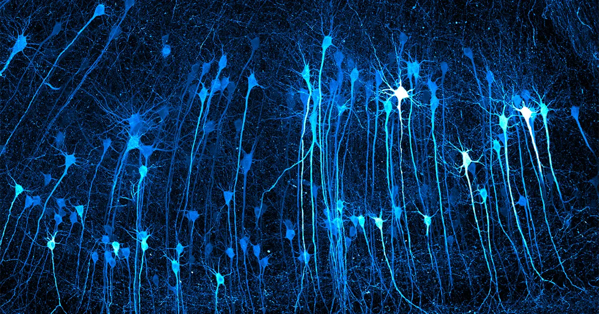
Nikon's Small World competition, established in 1974, continues to honor excellence in microscopy. This year's entries showcase the latest advancements, like confocal laser scanning microscopy, which illuminates thin layers of samples to build detailed images. Researchers use fluorescent dyes to label specific cellular structures, revealing mitochondria's dynamic roles in signaling and interactions with other organelles. This technology not only enhances our understanding of cellular processes but also highlights the importance of imaging in medical research. The competition's 49-year history reflects the evolution of microscopy and its impact on scientific discovery.
Key Points
Since 1974, Nikon's Small World competition honors microscopy achievements, celebrating 49 years of imaging technology.
Confocal laser scanning microscopy illuminates thin layers of samples, building detailed images line by line.
Fluorescent dyes label specific cellular structures, revealing mitochondria's dynamic roles in signaling and interactions.
Mitochondria are more dynamic than previously thought, likely involved in cytoplasmic streaming and cellular communication.
The competition highlights the evolution of microscopy and its impact on medical research and scientific discovery.
Why It Matters
The Nikon Small World competition showcases how imaging technology advances our understanding of cellular processes. Confocal laser scanning microscopy reveals mitochondria's dynamic roles, providing quantitative information about cellular structure and function. Researchers can use this technology to study cellular processes and develop new treatments for diseases.
Comments
Be the first to comment
Enjoyed this article?
Get it daily. 7am. Free. Reads in 5 minutes.